<< Chapter < Page Chapter >> Page >
By the end of this section, you will be able to:
  • Demonstrate the anatomical position
  • Describe the human body using directional and regional terms
  • Identify three planes most commonly used in the study of anatomy
  • Distinguish between the posterior (dorsal) and the anterior (ventral) body cavities, identifying their subdivisions and representative organs found in each
  • Describe serous membrane and explain its function

Anatomists and health care providers use terminology that can be bewildering to the uninitiated. However, the purpose of this language is not to confuse, but rather to increase precision and reduce medical errors. For example, is a scar “above the wrist” located on the forearm two or three inches away from the hand? Or is it at the base of the hand? Is it on the palm-side or back-side? By using precise anatomical terminology, we eliminate ambiguity. Anatomical terms derive from ancient Greek and Latin words. Because these languages are no longer used in everyday conversation, the meaning of their words does not change.

Anatomical terms are made up of roots, prefixes, and suffixes. The root of a term often refers to an organ, tissue, or condition, whereas the prefix or suffix often describes the root. For example, in the disorder hypertension, the prefix “hyper-” means “high” or “over,” and the root word “tension” refers to pressure, so the word “hypertension” refers to abnormally high blood pressure.

Anatomical position

To further increase precision, anatomists standardize the way in which they view the body. Just as maps are normally oriented with north at the top, the standard body “map,” or anatomical position    , is that of the body standing upright, with the feet at shoulder width and parallel, toes forward. The upper limbs are held out to each side, and the palms of the hands face forward as illustrated in [link] . Using this standard position reduces confusion. It does not matter how the body being described is oriented, the terms are used as if it is in anatomical position. For example, a scar in the “anterior (front) carpal (wrist) region” would be present on the palm side of the wrist. The term “anterior” would be used even if the hand were palm down on a table.

Regions of the human body

This illustration shows an anterior and posterior view of the human body. The cranial region encompasses the upper part of the head while the facial region encompasses the lower half of the head beginning below the ears. The eyes are referred to as the ocular region. The cheeks are referred to as the buccal region. The ears are referred to as the auricle or otic region. The nose is referred to as the nasal region. The chin is referred to as the mental region. The neck is referred to as the cervical region. The trunk of the body contains, from superior to inferior, the thoracic region encompassing the chest, the mammary region encompassing each breast, the abdominal region encompassing the stomach area, the coxal region encompassing the belt line, and the pubic region encompassing the area above the genitals. The umbilicus, or naval, is located at the center of the abdomen. The pelvis and legs contain, from superior to inferior, the inguinal or groin region between the legs and the genitals, the pubic region surrounding the genitals, the femoral region encompassing the thighs, the patellar region encompassing the knee, the crural region encompassing the lower leg, the tarsal region encompassing the ankle, the pedal region encompassing the foot and the digital/phalangeal region encompassing the toes. The great toe is referred to as the hallux. The regions of the upper limbs, from superior to inferior, are the axillary region encompassing the armpit, the brachial region encompassing the upper arm, the antecubital region encompassing the front of the elbow, the antebrachial region encompassing the forearm, the carpal region encompassing the wrist, the palmar region encompassing the palm, and the digital/phalangeal region encompassing the fingers. The thumb is referred to as the pollux. The posterior view contains, from superior to inferior, the cervical region encompassing the neck, the dorsal region encompassing the upper back and the lumbar region encompassing the lower back. The regions of the back of the arms, from superior to inferior, include the cervical region encompassing the neck, acromial region encompassing the shoulder, the brachial region encompassing the upper arm, the olecranal region encompassing the back of the elbow, the antebrachial region encompasses the back of the arm, and the manual region encompassing the palm of the hand. The posterior regions of the legs, from superior to inferior, include the gluteal region encompassing the buttocks, the femoral region encompassing the thigh, the popliteus region encompassing the back of the knee, the sural region encompassing the back of the lower leg, and the plantar region encompassing the sole of the foot. Some regions are combined into larger regions. These include the trunk, which is a combination of the thoracic, mammary, abdominal, naval, and coxal regions. The cephalic region is a combination of all of the head regions. The upper limb region is a combination of all of the arm regions. The lower limb region is a combination of all of the leg regions.
The human body is shown in anatomical position in an (a) anterior view and a (b) posterior view. The regions of the body are labeled in boldface.

A body that is lying down is described as either prone or supine. Prone describes a face-down orientation, and supine    describes a face up orientation. These terms are sometimes used in describing the position of the body during specific physical examinations or surgical procedures.

Regional terms

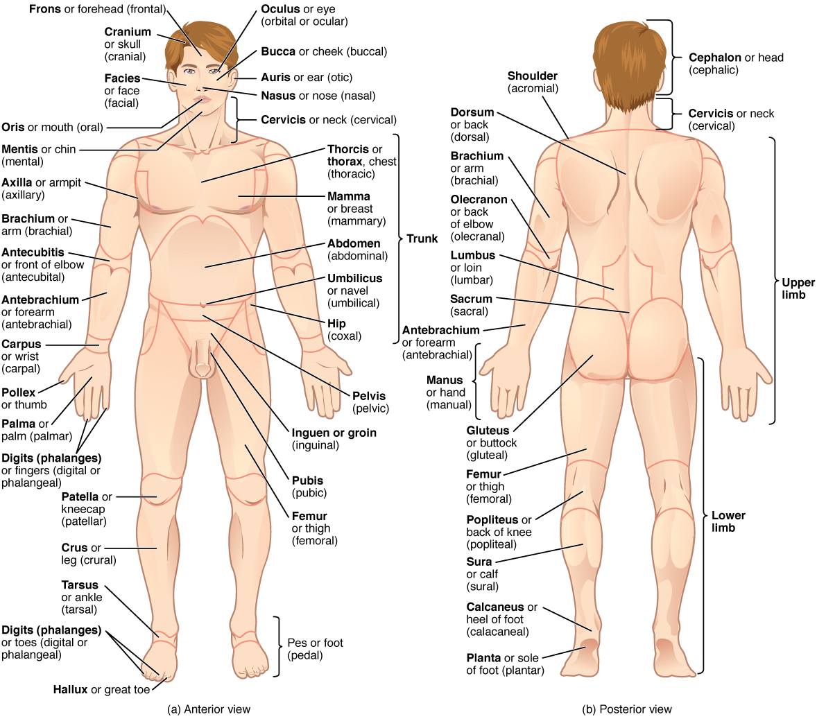
The human body’s numerous regions have specific terms to help increase precision (see [link] ). Notice that the term “brachium” or “arm” is reserved for the “upper arm” and “antebrachium” or “forearm” is used rather than “lower arm.” Similarly, “femur” or “thigh” is correct, and “leg” or “crus” is reserved for the portion of the lower limb between the knee and the ankle. You will be able to describe the body’s regions using the terms from the figure.

Questions & Answers

what is biology
Hajah Reply
the study of living organisms and their interactions with one another and their environments
AI-Robot
what is biology
Victoria Reply
HOW CAN MAN ORGAN FUNCTION
Alfred Reply
the diagram of the digestive system
Assiatu Reply
allimentary cannel
Ogenrwot
How does twins formed
William Reply
They formed in two ways first when one sperm and one egg are splited by mitosis or two sperm and two eggs join together
Oluwatobi
what is genetics
Josephine Reply
Genetics is the study of heredity
Misack
how does twins formed?
Misack
What is manual
Hassan Reply
discuss biological phenomenon and provide pieces of evidence to show that it was responsible for the formation of eukaryotic organelles
Joseph Reply
what is biology
Yousuf Reply
the study of living organisms and their interactions with one another and their environment.
Wine
discuss the biological phenomenon and provide pieces of evidence to show that it was responsible for the formation of eukaryotic organelles in an essay form
Joseph Reply
what is the blood cells
Shaker Reply
list any five characteristics of the blood cells
Shaker
lack electricity and its more savely than electronic microscope because its naturally by using of light
Abdullahi Reply
advantage of electronic microscope is easily and clearly while disadvantage is dangerous because its electronic. advantage of light microscope is savely and naturally by sun while disadvantage is not easily,means its not sharp and not clear
Abdullahi
cell theory state that every organisms composed of one or more cell,cell is the basic unit of life
Abdullahi
is like gone fail us
DENG
cells is the basic structure and functions of all living things
Ramadan
What is classification
ISCONT Reply
is organisms that are similar into groups called tara
Yamosa
in what situation (s) would be the use of a scanning electron microscope be ideal and why?
Kenna Reply
A scanning electron microscope (SEM) is ideal for situations requiring high-resolution imaging of surfaces. It is commonly used in materials science, biology, and geology to examine the topography and composition of samples at a nanoscale level. SEM is particularly useful for studying fine details,
Hilary
cell is the building block of life.
Condoleezza Reply
Got questions? Join the online conversation and get instant answers!
Jobilize.com Reply

Get Jobilize Job Search Mobile App in your pocket Now!

Get it on Google Play Download on the App Store Now


Source:  OpenStax, Anatomy & Physiology. OpenStax CNX. Feb 04, 2016 Download for free at http://legacy.cnx.org/content/col11496/1.8
Google Play and the Google Play logo are trademarks of Google Inc.

Notification Switch

Would you like to follow the 'Anatomy & Physiology' conversation and receive update notifications?

Ask