<< Chapter < Page Chapter >> Page >
By the end of this section, you will be able to:
  • Identify the axial muscles of the face, head, and neck
  • Identify the movement and function of the face, head, and neck muscles

The skeletal muscles are divided into axial    (muscles of the trunk and head) and appendicular    (muscles of the arms and legs) categories. This system reflects the bones of the skeleton system, which are also arranged in this manner. The axial muscles are grouped based on location, function, or both. Some of the axial muscles may seem to blur the boundaries because they cross over to the appendicular skeleton. The first grouping of the axial muscles you will review includes the muscles of the head and neck, then you will review the muscles of the vertebral column, and finally you will review the oblique and rectus muscles.

Muscles that create facial expression

The origins of the muscles of facial expression are on the surface of the skull (remember, the origin of a muscle does not move). The insertions of these muscles have fibers intertwined with connective tissue and the dermis of the skin. Because the muscles insert in the skin rather than on bone, when they contract, the skin moves to create facial expression ( [link] ).

Muscles of facial expression

The left panel in this figure shows the anterior view of the facial muscles, and the right panel shows the lateral view.
Many of the muscles of facial expression insert into the skin surrounding the eyelids, nose and mouth, producing facial expressions by moving the skin rather than bones.

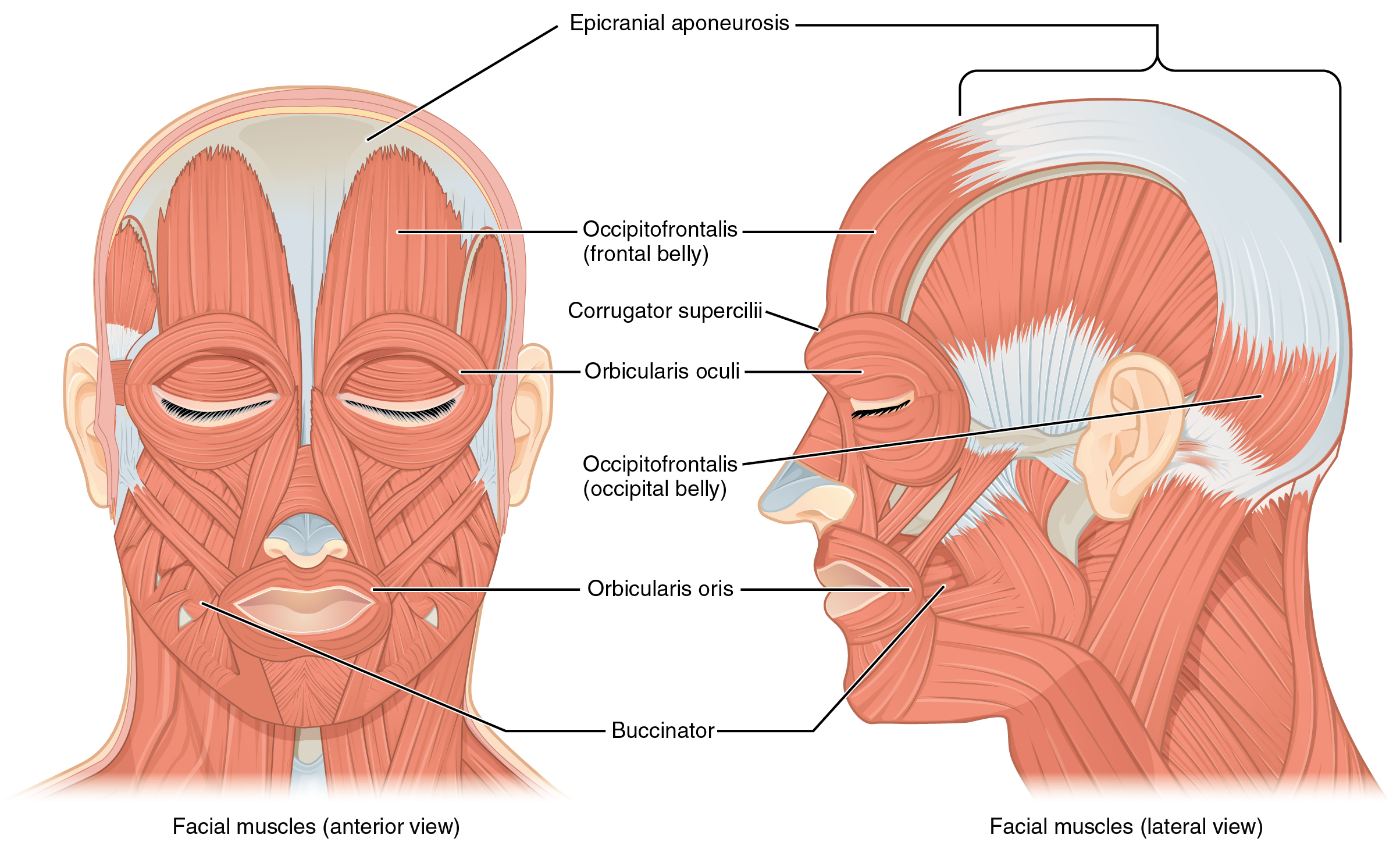
The orbicularis oris    is a circular muscle that moves the lips, and the orbicularis oculi    is a circular muscle that closes the eye. The occipitofrontalis    muscle moves up the scalp and eyebrows. The muscle has a frontal belly and an occipital (near the occipital bone on the posterior part of the skull) belly. In other words, there is a muscle on the forehead ( frontalis    ) and one on the back of the head ( occipitalis    ), but there is no muscle across the top of the head. Instead, the two bellies are connected by a broad tendon called the epicranial aponeurosis    , or galea aponeurosis (galea = “apple”). The physicians originally studying human anatomy thought the skull looked like an apple.

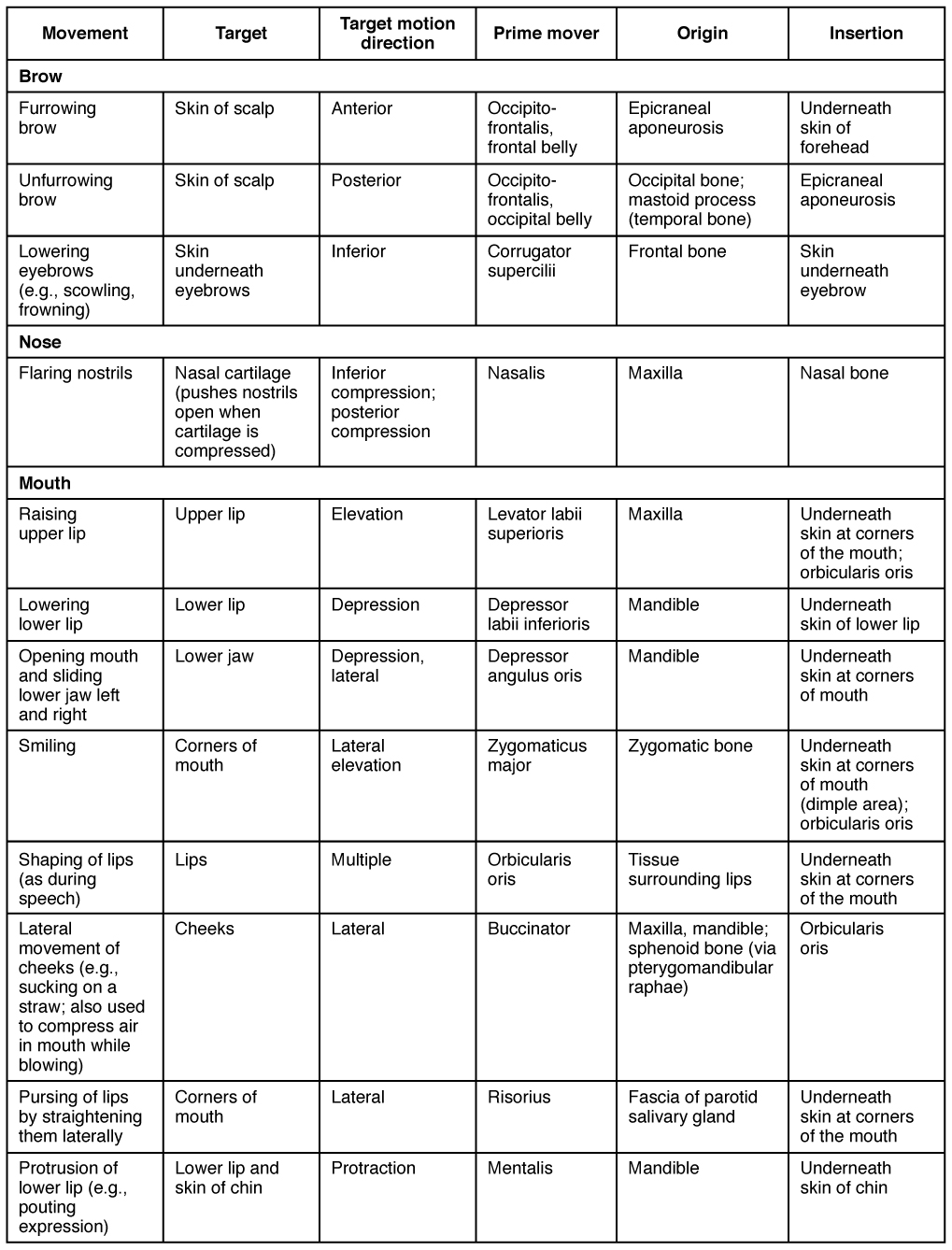
The majority of the face is composed of the buccinator    muscle, which compresses the cheek. This muscle allows you to whistle, blow, and suck; and it contributes to the action of chewing. There are several small facial muscles, one of which is the corrugator supercilii    , which is the prime mover of the eyebrows. Place your finger on your eyebrows at the point of the bridge of the nose. Raise your eyebrows as if you were surprised and lower your eyebrows as if you were frowning. With these movements, you can feel the action of the corrugator supercilli. Additional muscles of facial expression are presented in [link] .

Muscles in facial expression

This table describes the muscles used in facial expressions. To furrow the brow, the skin of the scalp moves in an anterior direction. The prime mover is the occipitofrontalis frontal belly, which originates from the epicraneal aponeurosis and inserts underneath the skin of the forehead. To unfurrow the brow, the skin of the scalp moves in the posterior direction. The prime mover is the occipitofrontalis occipital belly, which originates from the occipital bone and the mastoid process of the temporal bone and inserts into the epicraneal aponeurosis. To lower the eyebrows, as when scowling or frowning, the skin underneath the eyebrows moves in an inferior direction. The prime mover is the corrugator supercilii, which originates from the frontal bone and inserts into the skin underneath the eyebrow. To flare the nostrils, the nasal cartilage is compressed in an inferior and posterior direction. The prime mover is the nasalis, which originates from the maxilla and inserts into the nasal bone. Raising the upper lip involves elevating the upper lip tissue. The prime mover is the levator labii superioris, which originates from the maxilla and inserts underneath the skin at the corners of the mouth and also into the orbicularis oris. Lowering the lower lip involves depressing the lip and also moving it laterally. The prime mover is the depressor angulus oris, which originates from the mandible and inserts underneath the skin of the lower lip. Opening the mouth and sliding the lower jaw left and right involves depressing the lower jaw and also moving it laterally. The prime mover is thecdepressor angulus oris, which originates from the mandible and inserts underneath the skin at the corners of the mouth. Smiling involves elevating the corners of the mouth and also moving them in a lateral direction. The prime mover is the zygomaticus major, which originates from the zygomatic bone and inserts underneath the skin at the corners of the mouth in the dimple area, and also into the orbicularis oris. Shaping of the lips as during speech involves moving the lips in multiple directions. The prime mover is the orbicularis oris which originates from the tissue surrounding the lips and inserts underneath the skin at the corners of the mouth. Lateral movement of the cheeks such as when sucking on a straw or to compress air in the mouth while blowing involves moving the cheeks in a lateral direction. The prime mover is the buccinator, which originates from the maxilla, the mandible, and the sphenoid bone via the pterygomandibular raphae, and inserts into the orbicularis oris. Pursing of the lips by straightening them laterally involves moving the corners of the mouth in a lateral direction. The prime mover is the risorius, which originates from the fascia of the parotid salivary gland and inserts underneath the skin at the corners of the mouth. Protrusion of the lower lip, as when making a pouting expression, involves protracting the lower lip and the skin of the chin. The prime mover is the mentalis, which originates from the mandible and inserts underneath the skin of the chin.

Muscles that move the eyes

The movement of the eyeball is under the control of the extrinsic eye muscles    , which originate outside the eye and insert onto the outer surface of the white of the eye. These muscles are located inside the eye socket and cannot be seen on any part of the visible eyeball ( [link] and [link] ). If you have ever been to a doctor who held up a finger and asked you to follow it up, down, and to both sides, he or she is checking to make sure your eye muscles are acting in a coordinated pattern.

Questions & Answers

what is biology
Hajah Reply
the study of living organisms and their interactions with one another and their environments
AI-Robot
what is biology
Victoria Reply
HOW CAN MAN ORGAN FUNCTION
Alfred Reply
the diagram of the digestive system
Assiatu Reply
allimentary cannel
Ogenrwot
How does twins formed
William Reply
They formed in two ways first when one sperm and one egg are splited by mitosis or two sperm and two eggs join together
Oluwatobi
what is genetics
Josephine Reply
Genetics is the study of heredity
Misack
how does twins formed?
Misack
What is manual
Hassan Reply
discuss biological phenomenon and provide pieces of evidence to show that it was responsible for the formation of eukaryotic organelles
Joseph Reply
what is biology
Yousuf Reply
the study of living organisms and their interactions with one another and their environment.
Wine
discuss the biological phenomenon and provide pieces of evidence to show that it was responsible for the formation of eukaryotic organelles in an essay form
Joseph Reply
what is the blood cells
Shaker Reply
list any five characteristics of the blood cells
Shaker
lack electricity and its more savely than electronic microscope because its naturally by using of light
Abdullahi Reply
advantage of electronic microscope is easily and clearly while disadvantage is dangerous because its electronic. advantage of light microscope is savely and naturally by sun while disadvantage is not easily,means its not sharp and not clear
Abdullahi
cell theory state that every organisms composed of one or more cell,cell is the basic unit of life
Abdullahi
is like gone fail us
DENG
cells is the basic structure and functions of all living things
Ramadan
What is classification
ISCONT Reply
is organisms that are similar into groups called tara
Yamosa
in what situation (s) would be the use of a scanning electron microscope be ideal and why?
Kenna Reply
A scanning electron microscope (SEM) is ideal for situations requiring high-resolution imaging of surfaces. It is commonly used in materials science, biology, and geology to examine the topography and composition of samples at a nanoscale level. SEM is particularly useful for studying fine details,
Hilary
cell is the building block of life.
Condoleezza Reply
Got questions? Join the online conversation and get instant answers!
Jobilize.com Reply

Get Jobilize Job Search Mobile App in your pocket Now!

Get it on Google Play Download on the App Store Now


Source:  OpenStax, Anatomy & Physiology. OpenStax CNX. Feb 04, 2016 Download for free at http://legacy.cnx.org/content/col11496/1.8
Google Play and the Google Play logo are trademarks of Google Inc.

Notification Switch

Would you like to follow the 'Anatomy & Physiology' conversation and receive update notifications?

Ask