

| << Chapter < Page | Chapter >> Page > |
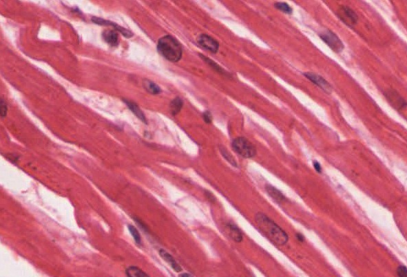
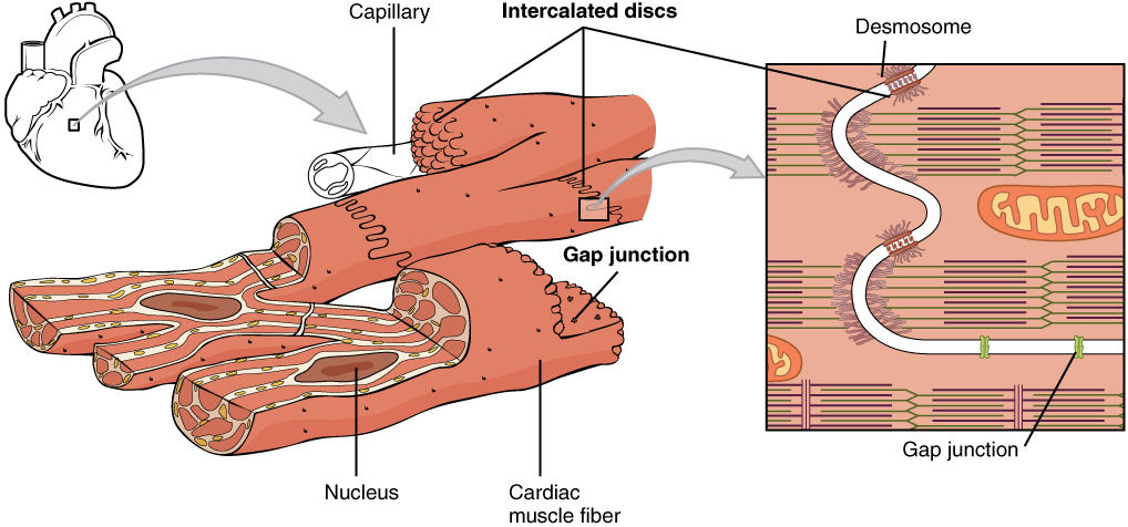
Cardiac muscle tissue is only found in the heart. Highly coordinated contractions of cardiac muscle pump blood into the vessels of the circulatory system. Similar to skeletal muscle, cardiac muscle is striated and organized into sarcomeres, possessing the same banding organization as skeletal muscle ( [link] ). However, cardiac muscle fibers are shorter than skeletal muscle fibers and usually contain only one nucleus, which is located in the central region of the cell. Cardiac muscle fibers also possess many mitochondria and myoglobin, as ATP is produced primarily through aerobic metabolism. Cardiac muscle fibers cells also are extensively branched and are connected to one another at their ends by intercalated discs. An intercalated disc allows the cardiac muscle cells to contract in a wave-like pattern so that the heart can work as a pump.
View the University of Michigan WebScope at (External Link) to explore the tissue sample in greater detail.
Intercalated discs are part of the sarcolemma and contain two structures important in cardiac muscle contraction: gap junctions and desmosomes. A gap junction forms channels between adjacent cardiac muscle fibers that allow the depolarizing current produced by cations to flow from one cardiac muscle cell to the next. This joining is called electric coupling, and in cardiac muscle it allows the quick transmission of action potentials and the coordinated contraction of the entire heart. This network of electrically connected cardiac muscle cells creates a functional unit of contraction called a syncytium. The remainder of the intercalated disc is composed of desmosomes. A desmosome is a cell structure that anchors the ends of cardiac muscle fibers together so the cells do not pull apart during the stress of individual fibers contracting ( [link] ).
Contractions of the heart (heartbeats) are controlled by specialized cardiac muscle cells called pacemaker cells that directly control heart rate. Although cardiac muscle cannot be consciously controlled, the pacemaker cells respond to signals from the autonomic nervous system (ANS) to speed up or slow down the heart rate. The pacemaker cells can also respond to various hormones that modulate heart rate to control blood pressure.
The wave of contraction that allows the heart to work as a unit, called a functional syncytium, begins with the pacemaker cells. This group of cells is self-excitable and able to depolarize to threshold and fire action potentials on their own, a feature called autorhythmicity ; they do this at set intervals which determine heart rate. Because they are connected with gap junctions to surrounding muscle fibers and the specialized fibers of the heart’s conduction system, the pacemaker cells are able to transfer the depolarization to the other cardiac muscle fibers in a manner that allows the heart to contract in a coordinated manner.
Another feature of cardiac muscle is its relatively long action potentials in its fibers, having a sustained depolarization “plateau.” The plateau is produced by Ca ++ entry though voltage-gated calcium channels in the sarcolemma of cardiac muscle fibers. This sustained depolarization (and Ca ++ entry) provides for a longer contraction than is produced by an action potential in skeletal muscle. Unlike skeletal muscle, a large percentage of the Ca ++ that initiates contraction in cardiac muscles comes from outside the cell rather than from the SR.
Cardiac muscle is striated muscle that is present only in the heart. Cardiac muscle fibers have a single nucleus, are branched, and joined to one another by intercalated discs that contain gap junctions for depolarization between cells and desmosomes to hold the fibers together when the heart contracts. Contraction in each cardiac muscle fiber is triggered by Ca ++ ions in a similar manner as skeletal muscle, but here the Ca ++ ions come from SR and through voltage-gated calcium channels in the sarcolemma. Pacemaker cells stimulate the spontaneous contraction of cardiac muscle as a functional unit, called a syncytium.
Notification Switch
Would you like to follow the 'Anatomy & Physiology' conversation and receive update notifications?

|
|

|
|

|
|

|
|

|
|
|
|

|
|

|
|

|
|

|
|

|
|

|